 |
| Optical Microscope |
Most cells are too small to be seen with the nacked eye. Without microscopes, we should know very little about them. The photographs(called photomicrographs)are images of plant cells and animal cells seen through the micrographs. A lamp lights the specimen of cells which the observer views through two magnifying lenses. The magnification of the cell is worked out as:
Total magnification of cells = magnifying power of the eye piece lens x magnifying power of the objective lens.
Good quality optical microscopes can magnify cells up to 2000times (x 2000)their original size. At higher magnification, the miage is enlarged but less clear because together the eye piece lens and objective lens of the micrscope can not distinguish between cell structures lying side-by-side-the syructures blur together appearing as one fuzzy image. The ability of amicroscope to distinguish between structures lying close together is called resolving power.
Instead of a beam of light, it passses a beam of electrons through the speciman of the cells. It can magnify uo to 1 million times (x 1000000), producing images which show the structures of cells in minute detail. Such detail is possible because the resolving power of the transmission electron microscope is much greater then that of the optical microscope.
 |
| Electron Microsocope |
One way to increase resolution is to increase magnification, so that small objects appear larger. Robert Hooke and Antonie van Leeuwenhoek were able to see small cells by magnifying their size, so that the cells appeared larger then the 100-micrometer limit imposed by human eye. Hooke and Leeuwenhoek accomplished this feat with microscopes that magnified images of cells by bending light through a glass lens. The size of the image that falls on the sheet of detector cells lining the back of your eye depends on how close the objects is to your eye - the closer the object, the bigger the image. Your eye, however, is incapable of focusing comfortably on an object closer then about 25 centimeters, because the eye is limited by the thickness of its lens.
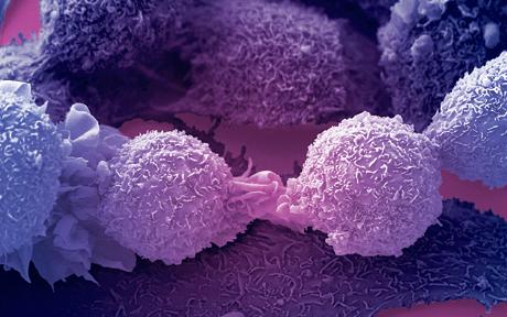 |
| Image of a cancer cell under microscope |
No comments:
Post a Comment